
BILDGEBUNG FÜR CHOLEZYSTEKTOMIEN
Entdecken Sie die klinischen Vorteile der aktiven Bildgebung mit intraoperativem Ultraschall für die laparoskopische Cholezystektomie. Unter Einsatz von bkActiv und dem erweiterten laparoskopischem Schallkopf können Ärzte:innen während des Eingriffs so oft wie nötig die Anatomie visualisieren und den Fortschritt überwachen.
Erfahren Sie in diesem Video, wie es funktioniert.
THE VALUE OF iUS IN CHOLECYSTECTOMIES
iUS is beneficial for cholecystectomies due to its safety, quick and repeatable imaging capabilities and ability to detect choledocholithiasis and enable visualization of biliary tract anatomy.
When helping to detect choledocholithiasis, iUS resulted in a 90-96% sensitivity rate and 100% specificity rate.7
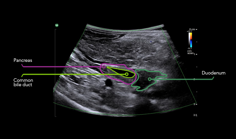
iUS is highly effective in visualizing biliary anatomy, which can be abnormal in as high as 40% of patient cases.8
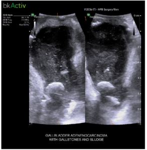
Gallbladder Adenocarcinoma with Gallstones and Sludge
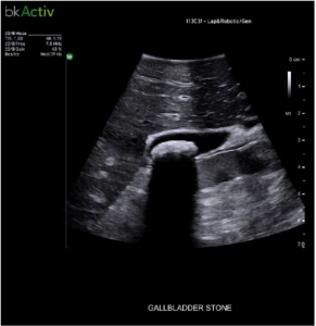
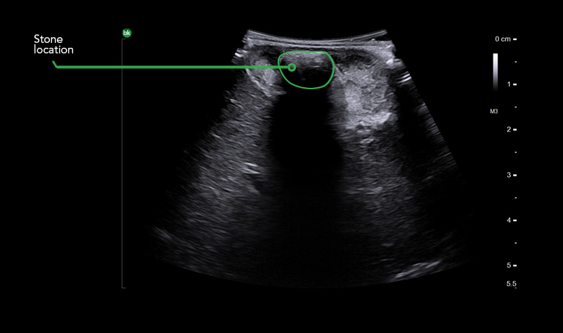
Gallstone

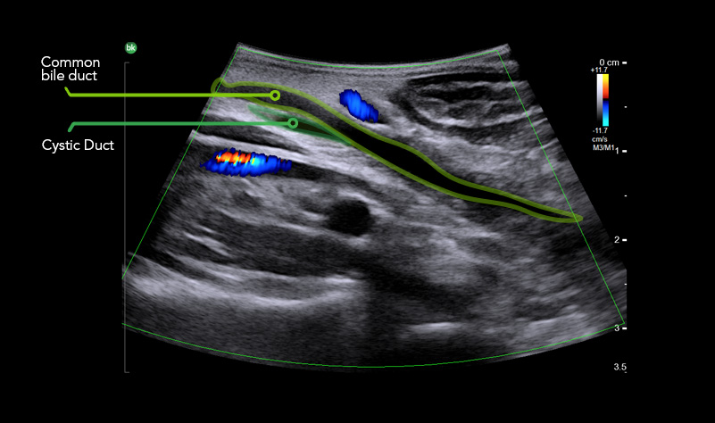
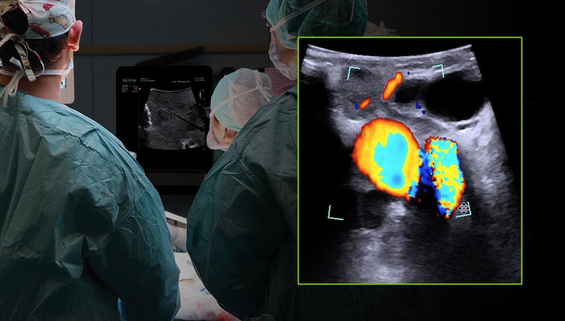
Bile Duct – Metastatic Tumor With Color Doppler
iUS is comparable to intraoperative cholangiography for imaging cholecystectomies.9
Learn more by watching our Laparoscopic Cholecystectomy Academy learning modules.
FALLSTUDIE
Diese Fallstudie zeigt die einzelnen Schritte bei der Nutzung von intraoperativem Ultraschall während eines laparoskopischen Cholezystektomie-Eingriffs, der von Dr. Somaiah Aroori, MB BS, MS (Surg), FRCS (Gen Surg), einem Berater für hepatobiliäre Chirurgie und Chirurgen für Nierentransplantation am Derriford Hospital and University Hospital Plymouth im Vereinigten Königreich durchgeführt wurde. Im Laufe des gesamten Eingriffs lieferte bkActiv Echtzeit-Informationen, die bei der präoperativen Bildgebung nicht erhältlich waren.
ADVANCED LAPAROSCOPIC TRANSDUCER I13C3f
Bietet eine hochauflösende Bildgebung für eine klare Visualisierung der Anatomie, mit flexibler 4-Weg-Spitze, die einen guten Organkontakt und Zugang zu schwer zugänglichen Bereichen ermöglicht, einer Laserfunktion, die ideal für die Visualisierung des Nadelverlaufs ist, und speziell entwickelten Nadelführungskanälen.
Laden Sie die PDF-Datei über aktive Bildgebung für biliäre Eingriffe herunter.
Sprechen Sie mit uns über aktive Bildgebung für Cholezystektomien.
-
1. Machi, J. (2009). The routine use of laparoscopic ultrasound decreases bile duct injury: a multicenter study. Surgical Endoscopy Journal, 23, 384-388.
2.Jamal, KN. (2016). Meta-analysis of the diagnostic accuracy of laparoscopic ultrasonography and intraoperative cholangiography in detection of common bile
duct stones. Royal college of surgeons, 98, 244-249.
3.Recommendation for cholecystectomy protocol based on intraoperative ultrasound – a single-centre retrospective case-control study (nih.gov)
4.Dili, A. (2017). Laparoscopic ultrasonography as an alternative to intraoperative cholangiography during laparoscopic cholecystectomy. World Journal of
Gastroenterology, 23, 5438-5450.
5.Intra-operative and laparoscopic ultrasound - Surgical Treatment - NCBI Bookshelf (nih.gov)
6.Intraoperative Sonography of the Biliary System : American Journal of Roentgenology : Vol. 177, No. 2 (AJR) (ajronline.org
7.https://www.sages.org/publications/guidelines/guidelines-for-the-use-of-laparoscopic-ultrasound/
8.Laparoscopic intraoperative biliary ultrasonography: findings during laparoscopic cholecystectomy for acute disease - PubMed (nih.gov)
9. https://www.sages.org/publications/guidelines/guidelines-for-the-use-of-laparoscopic-ultrasound/
*Where minimal time equates to ~5 minutes. The information included is meant to support the benefits of iUS as an imaging modality and is based on available literature. This is not product specific.